
KS3 Biology · Cells & life processes
Microscopes
- The parts of a light microscope and the job of each one
- How to focus, step by step, starting with the lowest power lens
- What a higher power lens does to the image, and how to carry a microscope
- How to make a scientific drawing, and what an electron microscope can show
- 2 min read
- 5 quick questions
- Free homework sheet
- Years 7 to 9
Revision notes
A light microscope shines light up through a thin specimen and uses two lenses to make an image that looks bigger. Total magnification = eyepiece × objective.
| Part | Job |
|---|---|
| Eyepiece lens | The lens you look through. It magnifies the image again, usually ×10. |
| Body tube | Holds the eyepiece the right distance above the objective lens, so the two lenses line up. |
| Nosepiece | Turns to click a different objective lens into place. |
| Objective lens | The lenses nearest the slide, usually ×4, ×10 and ×40. Always start with the lowest. |
| Stage clips | Hold the slide still on the stage. |
| Stage | The platform the slide sits on, with a hole for light to come up through. |
| Diaphragm | Controls how much light reaches the specimen. |
| Light source | Shines light up through the specimen. Older microscopes use a mirror instead. |
| Arm | Joins the top of the microscope to the base. Carry the microscope by the arm. |
| Coarse focusing wheel | Each turn moves the stage a long way. Use it on low power to get the image roughly in focus. |
| Fine focusing wheel | Each turn moves the stage a tiny way, for a sharp image. The only focusing wheel to use on high power. |
| Base | The heavy bottom that keeps it steady. Support it with your other hand when you carry it. |
How to focus:
- Put the slide on the stage, hold it with the clips and switch the light on.
- Turn the nosepiece to the lowest power objective lens.
- Looking from the side, use the coarse wheel to bring the stage up close to the lens.
- Look through the eyepiece and turn the coarse wheel slowly, moving the stage down, until the image appears.
- Use the fine focusing wheel to make the image sharp.
- Switch to a higher power objective lens and refocus with the fine wheel only.
Focusing makes the image clearer. A higher power lens makes it bigger, shows a smaller area and lets less light through. The image is upside down and back to front. Carry the microscope by the arm, with a hand under the base.
Scientific drawings: a sharp pencil; smooth, unbroken lines; no shading or colouring; labels with straight lines drawn with a ruler; a title and the magnification.
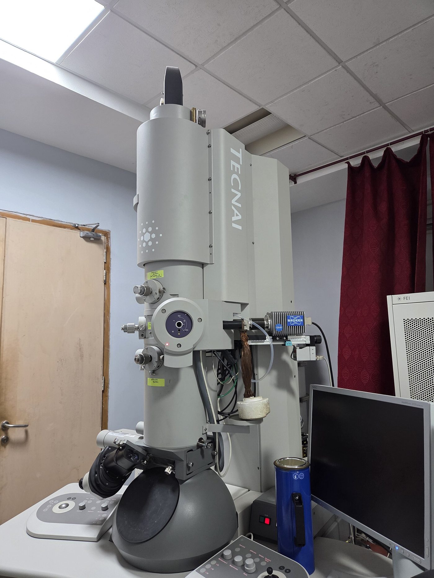
Stretch A light microscope shows cells, nuclei and chloroplasts. Ribosomes and viruses are far too small: an electron microscope magnifies over a million times, but it is huge, expensive, and the specimen must be dead.

Written to the KS3 national curriculum. Last changed 2 November 2026.
Key words
Say them, spell them, use them in an answer.
- light microscope
- How to focus
- clearer
- bigger
- smaller area
- Scientific drawings
- electron microscope
Common misconception
Often said“Turning the focusing wheel makes the image bigger.”
In factFocusing makes the image clearer. A higher power objective lens makes it bigger, and shows a smaller area.

Try 5 questions
Quick questions on microscopes, marked as you go. Each one explains the answer.
Question 1 of 5
Why does the stage have a hole in the middle?
Choose an answer. Each question explains itself.
The homework sheet
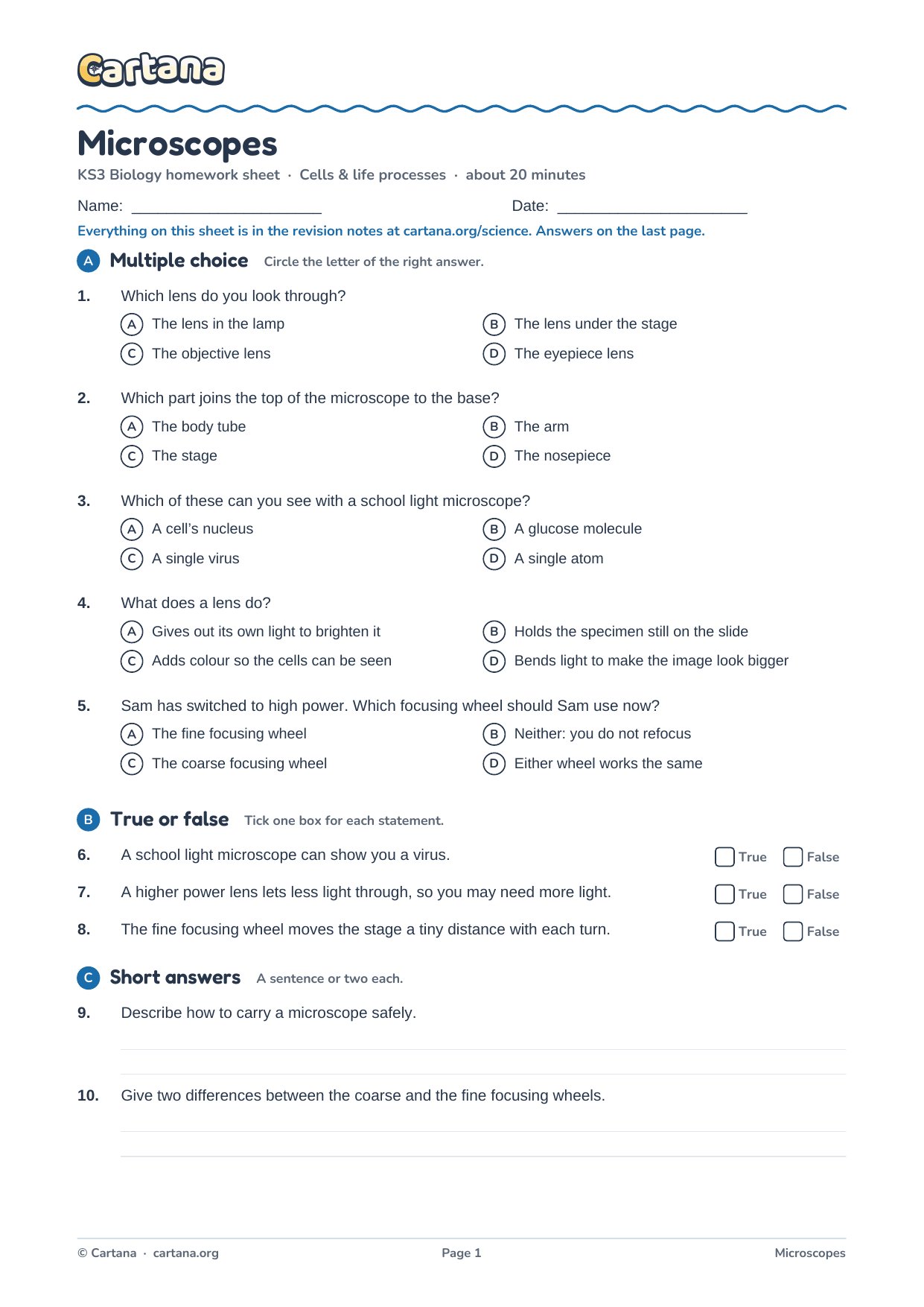
About twenty minutes. Everything on it is in the notes above; the answers are on the last page.
Free homework sheet
Microscopes homework sheet
17 questions, about twenty minutes, with the answers on the last page: multiple choice, true or false, short answers, key words to define and one to explain in full.
KS3 Cells & life processes
Revised the whole unit? There is a 40-mark Cells & life processes test paper with a mark scheme.
 Watch the trailer · 1:06
New from Cartana · Beta test 1–31 December
Revision that feels like an adventure
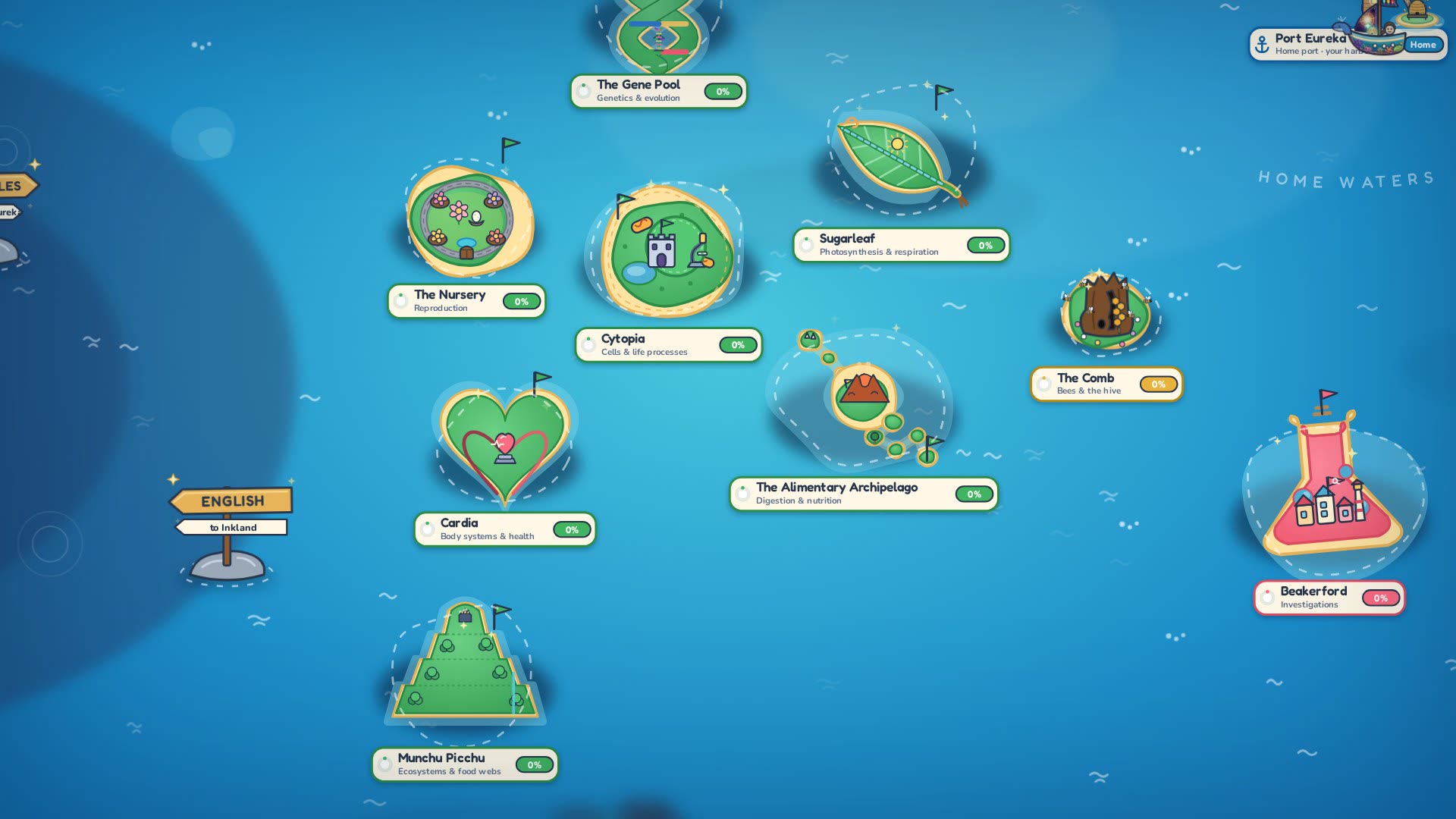
Cartana is our KS3 learning adventure: sail between islands, answer quick-fire questions and battle bosses. It starts with science, with maths on the way, and launches in 2027. This December a small group of testers gets to play it first.
In the Cartana adventure, microscopes is The Lens Vault, a district of the island Cytopia.
- Play the game before its official release
- Help us build it with your feedback
- Earn exclusive in-game items in return
More in this unit